This informal CPD article ‘Muscle Shortening and Joint Dysfunction: a Physical-Clinical Explanation’ was provided by Dr. Mauro Lastrico, Physioterapist at AIFiMM Formazione, an organisation recognised by the Italian Ministry of Health as an authoried CME provider. They offer organised training courses in the Mézières Method, a rehabilitative and postural approach.
Clinical Assessment of Muscle Shortening
In everyday clinical practice, it is often observed that, even in the absence of any specific pathology — congenital, acquired, or neurological — certain muscles progressively lose part of their resting length. The contractile capacity remains preserved, but the reduced baseline length modifies the geometric relationships between the articular segments, introducing mechanical constraints that were not present under physiological conditions. The most evident consequences are a decrease in articular range of motion (both active and passive) and a reorganisation of the normal movement sequence, with anticipations or delays of functional segments to compensate for the new resistance.
To understand the underlying mechanisms, it is useful to consider the problem from a mechanical point of view, examining the behaviour of the passive components of the musculotendinous system as viscoelastic materials[4-6]. It is within these tissues that the variations in stiffness we clinically recognise as "shortening" are generated and progressively accumulated.
The Physical Laws of Elastic Deformation of Materials
According to materials physics[1-3], every body subjected to a force deforms in proportion to the applied stress and its intrinsic elasticity. This principle also applies to biological tissues, which behave as viscoelastic materials, capable of deforming under load and only partially recovering their initial shape.
In a theoretical model, a perfectly elastic material (elasticity coefficient = 1) would fully restore the accumulated energy, returning exactly to the original state. In reality, however, all biological materials have a coefficient less than 1, which results in energy loss and residual deformation, even minimal, after each stress.
The magnitude of such deformation depends on two variables: the intensity of the force and the duration of its application. In general terms, the longer the time of force application, the greater the permanent deformation. The time variable, in fact, allows the material to reorganise its internal structure, generating stable adaptations.
This means that even moderate forces, if prolonged, can induce [18,19]significant structural modifications, while intense but short-duration loads tend to produce more limited deformations, although never completely reversible. This principle, valid for any material, offers the interpretative key to understanding the processes of mechanical adaptation and progressive shortening in muscle and connective tissues.
Application of Physical Laws to Muscle Tissue
In muscle, two types of elastic materials coexist with different mechanical characteristics:
- the contractile component, formed by the actin and myosin filaments[7-9],
- the connective component, consisting of the membranes (endomysium, perimysium, epimysium), the aponeuroses, and the tendons.
The contractile part can only contract and relax: its elasticity coefficient is high and allows an almost complete recovery of the initial length after each contraction. For this reason, alterations affecting it manifest more frequently as variations in baseline tone or an increase in active tension, rather than as permanent structural modifications.
The connective component, on the other hand, has a lower elasticity coefficient and a behaviour more typical of viscoelastic materials: under repeated or prolonged mechanical stimuli over time, it can maintain residual deformations[23-25] proportional to the intensity, duration, and frequency of the stresses sustained.
In practical terms, this means that persistent compression or traction can induce a stable shortening or lengthening of the connective matrix, consequently modifying the functional length of the muscle as a whole.
This difference in behaviour explains a recurring clinical observation:
- muscle relaxation or tone inhibition techniques act effectively on the contractile component, reducing baseline tension;
- conversely, shortenings structured over time — due to modifications of the connective tissue — respond in a limited way to such strategies, requiring more prolonged mechanical stimuli[26-28] to induce real remodelling.

Elastic elements of muscle
Mechanical Classification of Elastic Components
From a mechanical point of view, the elastic elements of muscle are divided into two main categories, defined based on their arrangement and behaviour under load:
- Series elastic elements[13-15]
- Parallel elastic elements[10,12]
1. Series Elastic Elements
They comprise the tendons and their extensions within the muscle belly. Their main function is to cushion the stresses generated during contraction and to return part of the accumulated energy at the end of the effort, analogously to a spring that stretches and then releases elastic energy.
During contraction, these elements deform in traction and, thanks to the presence of sensory structures such as the Golgi tendon organs[16,17], can trigger reflex mechanisms of protective relaxation when internal tension exceeds a safety threshold.
2. Parallel Elastic Elements
They consist of the sarcolemma, the connective membranes, and the connective tissue interposed between muscle fibres. Their task is to dampen the stresses generated by stretches, distributing the force and reducing internal resistances.
During contraction, these elements are compressed laterally: the internal pressure increases as a function of the product force × time. When such stimulus is prolonged, the connective components in parallel do not completely recover their original shape, leaving a portion of residual deformation. It is at this level that the structural shortening of muscle originates, that is, the progressive loss of baseline length observed clinically.
Physical Model of the Muscle Fibre
If we represent the behaviour of the muscle fibre in physical-mathematical terms, we can distinguish three phases:
- Rest — the muscle presents its physiological length, with equilibrium between the contractile and connective components.
- Contraction — the actin and myosin fibres actively shorten, generating force and transmitting traction to the connective components arranged in parallel. The latter are thus compressed proportionally to the intensity and duration of the contraction.
- Relaxation — once the stimulus ends, the contractile part, having high elasticity, tends to return to its initial length, but the connective component, having a lower elasticity coefficient, maintains part of the compressive deformation.
With the maintenance of contraction over time, the internal pressure on connective components increases progressively and may go beyond the tissue’s elastic recovery threshold. Therefore, it is prolonged or sustained contractions, more than short ones interspersed with release phases, that determine persistent compressive deformations and the reduction of the muscle's baseline length. When instead the contraction is repeated but followed by adequate relaxation time, the tissue has a greater recovery margin and the cumulative impact on connective structures is lower.
In summary, it is the duration of sustained tension — rather than the frequency of contraction — that determines the degree of residual shortening of muscle and the establishment of structural modifications in connective tissue.
The Skeletal Consequences of Muscle Shortening
Sustained muscle contractions with approximation of insertions, or isometric contractions not performed in maximum physiological lengthening, determine different effects on the two main components of muscle.
The contractile part — consisting of the actin and myosin filaments — tends to develop an increase in baseline tone, a consequence of its high elastic coefficient and of the neuromuscular adaptation to tension.
The connective part, on the other hand, possessing a lower elasticity coefficient, undergoes progressive permanent deformations proportional to the intensity and duration of the contraction, with a consequent reduction in baseline length.
At a functional level, this dual alteration determines an increase in resistance force (that is, the amount of energy that the muscle must use to overcome its own internal tensions) and, in parallel, a reduction in work capacity and mechanical power. The system therefore becomes less efficient: to produce the same movement requires more energy, and part of it is dissipated in overcoming the internal stiffness of the muscle itself.
The result of these modifications also manifests on the skeletal plane.
Bones, being passive structures, adapt to the muscular forces acting on them: when a shortened muscle exerts a constant traction, the articular segments are progressively drawn in the direction of the dominant vector.
A particularly evident clinical example is that of glenohumeral impingement[29-31]. In this case, the vector dominance of the internal rotator muscles of the humerus (such as the pectoralis major, latissimus dorsi, and teres major) over the external rotators (infraspinatus and teres minor) tends to internally rotate and anteriorise the humeral head, bringing it closer to the glenoid cavity.
This deviation of the traction vector alters articular centring and reduces the subacromial space, generating over time a mechanical impingement[29] between the humeral head and the overlying structures (supraspinatus tendon, subacromial bursa).
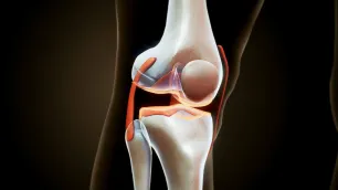
The phenomenon therefore does not arise from a "primary lesion" but from a progressive modification of force and length relationships between antagonistic muscle groups, which pushes the bony component towards a new geometric equilibrium. Similarly, in other body regions — such as the hip or knee — chronic asymmetric tensions can concentrate load vectors on limited areas of the articular surfaces, generating over time localised overload. In this sense, osteoarthritis is not so much the initial cause as a possible secondary mechanical consequence[35,36] of persistent muscular imbalance.[32-34]
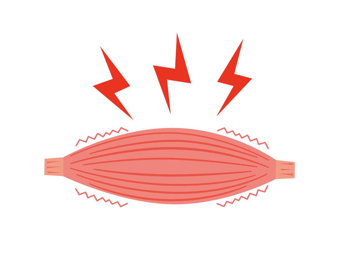
Sustained muscle contractions
The Relationship Between Resistance Force, Work, and Power
The progressive shortening of the connective component, associated with the increase in baseline tone of the contractile part, determines an increase in the resistance force of the muscle. By the term resistance force we mean the passive resistance that the muscle opposes to lengthening: an internal force that manifests when attempting to extend an articular segment. The increase in this resistance entails a consequent decrease in work capacity and power of the muscle.
Work is defined as the product of force and displacement[37,38]: it represents the amount of energy actually transformed into movement.
Power instead indicates the work produced per unit of time, that is, the dynamic efficiency with which the muscle is able to act.
In a shortened muscle, although the contractile capacity of the actin and myosin fibres remains intact, part of the developed energy must be used to overcome the internal resistances of the connective tissue before producing useful displacement.
The principle is analogous to that of a mechanical system with internal friction: the motor functions correctly, but part of its energy is dissipated to overcome internal friction, reducing the overall effectiveness of movement. It follows that the shortened muscle is not truly weak, but inefficient from a mechanical point of view. The reduction in performance does not derive from a loss of force, but from a non-optimal energy use, largely destined to overcome passive tensions rather than to generate movement.
This principle finds clear clinical confirmation in situations of prolonged immobilisation[20-22]. A typical example is the patient who, after removal of a cast for humerus fracture, presents the elbow in flexion. In this case, the flexor muscles have maintained for weeks a position of shortening, undergoing continuous compression. The connective tissue surrounding them has adapted to that condition, developing a greater resistance force.
Consequently, at the time of cast removal:
- the flexors oppose resistance to extension, both during passive lengthening induced by the therapist and during triceps activation;
- their dynamic capacity is reduced, since part of the energy generated by contraction is dissipated within the muscle itself.
This example highlights a crucial point: the differentiation between resistance force, work, and power is indispensable to understand how muscle shortening does not only alter articular statics, but also conditions the dynamics of movement and the overall efficiency of the system.
Concluding Considerations on the Operational Validity of the Model
Muscle shortening, understood as reduction of the available length of the muscle in the absence of specific pathology, is a constantly observable empirical datum in clinical practice. It is detectable through the evaluation of articular range of motion and lengthening tests, and represents a condition that the physiotherapist can measure, describe, and modify during treatment.
The hypothesis proposed in this article — according to which such shortening derives mainly from a viscoelastic deformation of the connective components induced by compressive forces prolonged over time — constitutes a coherent interpretative model, founded on the application of the principles of materials physics to biological tissues.
At present, a direct experimental verification of this mechanism at the histological level is not feasible: it would require analysis of the tissue in vivo during loading and release phases, a condition that is technically inaccessible. Even traditional mechanical tests, based on tests of traction of muscle tissue, do not faithfully reproduce the conditions of chronic compression that are established in vivo during sustained contractions or prolonged postures.
Consequently, the truth of this hypothesis should be understood on a clinical-operational plane, where the value of the model does not lie in direct demonstration, but in the ability to predict and explain observable phenomena.
In this sense:
- The model provides a coherent explanation of alterations in articular sequence, allowing us to predict vector dominances and the muscular compensations that derive from them.
- It finds indirect confirmation in the therapeutic results obtained with interventions aimed at recovering length of the connective component and rebalancing between Resistance Force and Work Force, which frequently lead to normalisation of articular vectors and resolution of symptoms.
The model, therefore, does not claim to describe a conclusive mechanism, but proposes itself as a causal hypothesis verifiable on the clinical plane. As long as observation shows that muscle shortening alters articular axes and that length recovery restores equilibrium and function, the model will maintain its operational validity.
In accordance with the epistemological thinking of Karl Popper[39,40], its solidity does not derive from a presumed certainty, but from the ability to resist refutation: each new clinical observation represents both a verification and a possible falsification. As long as clinical results continue to confirm its predictions, the biomechanical model of muscle shortening can be considered provisionally true, that is, scientifically useful in everyday practice.
We hope this article was helpful. For more information from AIFiMM Formazione, please visit their CPD Member Directory page. Alternatively, you can go to the CPD Industry Hubs for more articles, courses and events relevant to your Continuing Professional Development requirements.
REFERENCES
1. Timoshenko SP, Goodier JN. Theory of Elasticity. 3rd ed. New York: McGraw-Hill; 1970.
2. Beer FP, Johnston ER, DeWolf JT, Mazurek DF. Mechanics of Materials. 7th ed. New York: McGraw-Hill Education; 2015.
3. Landau LD, Lifshitz EM. Theory of Elasticity. 3rd ed. Oxford: Butterworth-Heinemann; 1986.
4. Fung YC. Biomechanics: Mechanical Properties of Living Tissues. 2nd ed. New York: Springer-Verlag; 1993.
5. Nordin M, Frankel VH. Basic Biomechanics of the Musculoskeletal System. 4th ed. Philadelphia: Lippincott Williams & Wilkins; 2012.
6. Humphrey JD. Continuum biomechanics of soft biological tissues. Proc R Soc Lond A. 2003;459(2029):3-46.
7. Lieber RL, Ward SR. Skeletal muscle design to meet functional demands. Philos Trans R Soc Lond B Biol Sci. 2011;366(1570):1466-1476.
8. Herzog W. Skeletal muscle mechanics: questions, problems and possible solutions. J Neuroeng Rehabil. 2017;14(1):98.
9. Lieber RL, Fridén J. Functional and clinical significance of skeletal muscle architecture. Muscle Nerve. 2000;23(11):1647-1666.
10. Purslow PP. The structure and role of intramuscular connective tissue in muscle function. Front Physiol. 2020;11:495.
11. Gillies AR, Lieber RL. Structure and function of the skeletal muscle extracellular matrix. Muscle Nerve. 2011;44(3):318-331.
12. Prado LG, Makarenko I, Andresen C, Krüger M, Opitz CA, Linke WA. Isoform diversity of giant proteins in relation to passive and active contractile properties of rabbit skeletal muscles. J Gen Physiol. 2005;126(5):461-480.
13. Hill AV. The heat of shortening and the dynamic constants of muscle. Proc R Soc Lond B Biol Sci. 1938;126(843):136-195.
14. Morgan DL. New insights into the behaviour of muscle during active lengthening. Biophys J. 1990;57(2):209-221.
15. Proske U, Morgan DL. Tendon stiffness: methods of measurement and significance for the control of movement. J Biomech. 1987;20(1):75-82.
16. Houk JC, Henneman E. Responses of Golgi tendon organs to active contractions of the soleus muscle of the cat. J Neurophysiol. 1967;30(3):466-481.
17. Jami L. Golgi tendon organs in mammalian skeletal muscle: functional properties and central actions. Physiol Rev. 1992;72(3):623-666.
18. Goldspink G, Tabary C, Tabary JC, Tardieu C, Tardieu G. Effect of denervation on the adaptation of sarcomere number and muscle extensibility to the functional length of the muscle. J Physiol. 1974;236(3):733-742.
19. Williams PE, Goldspink G. Changes in sarcomere length and physiological properties in immobilised muscle. J Anat. 1978;127(Pt 3):459-468.
20. Tabary JC, Tabary C, Tardieu C, Tardieu G, Goldspink G. Physiological and structural changes in the cat's soleus muscle due to immobilisation at different lengths by plaster casts. J Physiol. 1972;224(1):231-244.
21. Herbert RD, Crosbie J. Rest length and compliance of non-immobilised and immobilised rabbit soleus muscle and tendon. Eur J Appl Physiol. 1997;76(5):472-479.
22. Trudel G, Uhthoff HK. Contractures secondary to immobility: is the restriction articular or muscular? An experimental longitudinal study in the rat knee. Arch Phys Med Rehabil. 2000;81(1):6-13.
23. Woo SL, Gomez MA, Akeson WH. The time and history-dependent viscoelastic properties of the canine patellar tendon. J Biomech Eng. 1981;103(4):293-298.
24. Best TM, McElhaney J, Garrett WE Jr, Myers BS. Characterisation of the passive responses of live skeletal muscle using the quasi-linear theory of viscoelasticity. J Biomech. 1994;27(4):413-419.
25. Gajdosik RL. Passive extensibility of skeletal muscle: review of the literature with clinical implications. Clin Biomech. 2001;16(2):87-101.
26. Magnusson SP, Simonsen EB, Aagaard P, Kjaer M. Biomechanical responses to repeated stretches in human hamstring muscle in vivo. Am J Sports Med. 1996;24(5):622-628.
27. Weppler CH, Magnusson SP. Increasing muscle extensibility: a matter of increasing length or modifying sensation? Phys Ther. 2010;90(3):438-449.
28. Freitas SR, Mendes B, Le Sant G, Andrade RJ, Nordez A, Milanovic Z. Can chronic stretching change the muscle-tendon mechanical properties? A review. Scand J Med Sci Sports. 2018;28(3):794-806.
29. Michener LA, McClure PW, Karduna AR. Anatomical and biomechanical mechanisms of subacromial impingement syndrome. Clin Biomech. 2003;18(5):369-379.
30. Ludewig PM, Cook TM. Alterations in shoulder kinematics and associated muscle activity in people with symptoms of shoulder impingement. Phys Ther. 2000;80(3):276-291.
31. Cools AM, Declercq GA, Cambier DC, Mahieu NN, Witvrouw EE. Trapezius activity and intramuscular balance during isokinetic exercise in overhead athletes with impingement symptoms. Scand J Med Sci Sports. 2007;17(1):25-33.
32. Janda V. Muscles and motor control in cervicogenic disorders: assessment and management. In: Grant R, editor. Physical Therapy of the Cervical and Thoracic Spine. New York: Churchill Livingstone; 1994. p. 195-216.
33. Sahrmann SA. Diagnosis and Treatment of Movement Impairment Syndromes. St. Louis: Mosby; 2002.
34. Page P, Frank CC, Lardner R. Assessment and Treatment of Muscle Imbalance: The Janda Approach. Champaign: Human Kinetics; 2010.
35. Felson DT. Osteoarthritis as a disease of mechanics. Osteoarthritis Cartilage. 2013;21(1):10-15.
36. Andriacchi TP, Mündermann A. The role of ambulatory mechanics in the initiation and progression of knee osteoarthritis. Curr Opin Rheumatol. 2006;18(5):514-518.
37. Knudson D. Fundamentals of Biomechanics. 2nd ed. New York: Springer; 2007.
38. Winter DA. Biomechanics and Motor Control of Human Movement. 4th ed. Hoboken: John Wiley & Sons; 2009.
39. Popper KR. The Logic of Scientific Discovery. London: Hutchinson; 1959.
40. Popper KR. Conjectures and Refutations: The Growth of Scientific Knowledge. London: Routledge; 1963.