This informal CPD article ‘Vector Analysis of the Vertebral Column in the Sagittal Plane - Clinical Application to Vertebral Equilibrium’, was provided by Dr. Mauro Lastrico, Physiotherapist at AIFiMM Formazione, an organisation recognised by the Italian Ministry of Health as an authorised CME provider. They offer organised training courses in the Mézières Method, a rehabilitative and postural approach.
This article represents the fifth contribution in a series dedicated to applying the principles of physics to the musculoskeletal system. The first article, Clinical Assessment of Muscle Shortening [1], introduced the physical model of muscle shortening as viscoelastic deformation of connective components, distinguishing the mechanical behaviour of the contractile component from that of the connective component.
The second article, Body Equilibrium - A Physical-Clinical Interpretation of Human Upright Stability [2], presented the interpretation of human equilibrium as alignment between the weight force (G) and the ground reaction force (R), refuting the concept of "antigravity muscles".
The third and fourth articles completed the theoretical framework by introducing vector analysis as a tool for identifying the muscular causes of alterations in physiological joint sequence: Vector Analysis in Musculoskeletal Biomechanics - Part 1: Foundations and Clinical Principles [3] presented the fundamental principles of vector analysis, while Vector Analysis in Musculoskeletal Biomechanics - Part 2: Clinical Applications and Case Interpretation [4] illustrated its clinical applications.
The present work applies these principles to the analysis of the vertebral column in the sagittal plane, demonstrating how the distribution of muscular forces determines the configuration of physiological curves and how variations in these forces modify the articular sequence. The objective is not to propose an alternative paradigm to evidence-based physiotherapy, but to provide an interpretive tool that enables us to understand why certain joint alterations manifest with predictable patterns, and how to direct therapeutic intervention towards primary causes rather than secondary manifestations [3,4,5].
1. Introduction to Vector Analysis in the Sagittal Plane
The sagittal plane constitutes the reference framework for analysing the physiological curves of the spine and their equilibrium relationships. By identifying the dominant vectors and mechanical resultants underlying variations in the physiological curves of the vertebral sinusoid, district-level analysis maintains coherence with the general physical model, demonstrating how the same laws - tissue elasticity, force moments, equilibrium between G and R forces - find concrete expression in the human body [2,3,6].
Vector analysis enables us to identify the muscular dominances present in each tract and the mechanisms by which antagonist muscles attempt to balance dominant forces [3,6]. Analysis in the sagittal plane does not represent a two-dimensional reduction of system complexity, but rather an analytical tool that allows isolation of dominant vectorial components within three-dimensional behaviour [3,7].
1.1 Myofunctional Subdivision
Observing the anatomical disposition of muscles influencing the cranium, vertebral column and pelvis reveals that muscular insertions do not respect anatomical boundaries but create functional units that encompass multiple vertebral segments [8]. This observation leads to supplementing anatomical classification with a myofunctional subdivision distinguished into three tracts: cranio-cervico-dorsal lordosis (extending from the cranium to the spinous process of T3), dorsal kyphosis (extending from the spinous process of T4 to that of T6), and dorso-lumbo-sacral lordosis (extending from the spinous process of T7 to the sacrum).
The anatomical lordosis C1-C7 is muscularly prolonged to T3 through the action of specific muscle groups [8,9]. Posteriorly act the trapezius (upper fibres) inserting on the scapula, the levator scapulae connecting the cervical vertebrae to the scapula, and the paravertebral muscles. The superomedial angle of the scapula is positioned anatomically at the level of T3, creating a continuous functional unit. Anteriorly act the anterior paravertebral muscles (longus capitis, longus colli and rectus capitis anterior), the scalenes and the sternocleidomastoid muscles.
1.2 Biomechanical Function of the Curves
Functionally, the vertebral sinusoid serves to cushion vertical loads [10]. If the spine were straight it would suffer greater compressive damage from the direct effect of compression forces. The presence of curves transforms vertical compression forces into more manageable components, distributing loads throughout the entire structure [10,11].
The development of vertebral curves during growth confirms this functional vision [12]. The neonate presents the vertebral column in total kyphosis (primary curve). During growth, muscles between the cranium and T3 are used to create the cervico-dorsal lordosis that permits cranial control and horizontal vision. Subsequently, muscles between T7 and the sacrum are used to create the dorso-lumbo-sacral lordosis that enables achievement of upright posture [12,13]. This sequential development demonstrates how vertebral curves result from muscular action rather than simple anatomical adaptations.
In the following analysis, we will examine possible modifications of vertebral tracts caused by shortening of muscles acting on each functional unit. Every alteration in one of the three functional units can influence the others; muscular shortenings modify curves according to predictable patterns; symptomatology may manifest at a distance from the primary biomechanical cause [5,14].
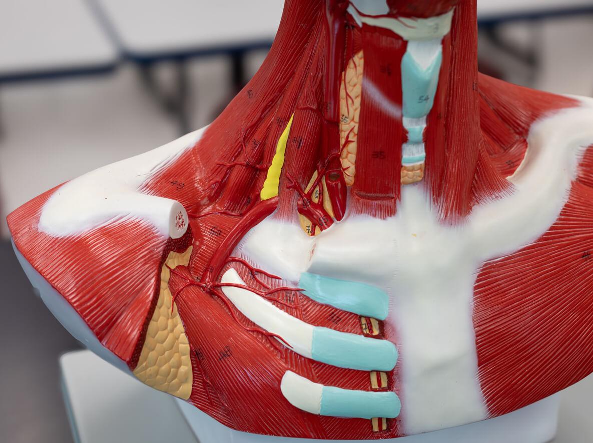
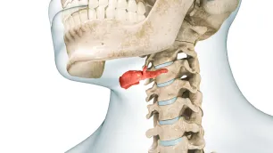
Anterior neck muscles
2. Cranio-Cervico-Dorsal Lordosis (C0-T3)
The cranio-cervico-dorsal tract presents a specific characteristic compared to the rest of the vertebral column: it is the only segment where paravertebral muscles with double insertion are present, both anteriorly (anterior neck muscles) and posteriorly (posterior paravertebral muscles) [8,9]. This anatomical peculiarity determines complex and particular biomechanical mechanisms for this district.
Anteriorly act the scalenes, sternocleidomastoid muscles and anterior neck muscles (rectus capitis anterior, longus capitis, longus colli). Posteriorly act the posterior paravertebral muscles, levator scapulae and upper fibres of the trapezius [9].
2.1 Vector Analysis of Posterior Muscles
Increased basal tone and subsequent shortening of posterior muscles determine two principal effects: posterior flexion of the cranium (through direct action of paravertebral muscles and upper fibres of trapezius, and indirect action of levator scapulae) and increased cervical lordosis (through direct action of paravertebral muscles and levator scapulae, and indirect action of upper fibres of trapezius) [3,9,15].
The upper fibres of trapezius flex the cranium posteriorly and, through mechanical resultant, increase vertebral lordosis. The levator scapulae flex posteriorly the vertebrae from C1 to C4 and increase cervical lordosis in toto. The paravertebral muscles flex the cranium posteriorly and increase cervico-dorsal lordosis from C1 to T3 [15,16].
2.2 Vector Analysis of Anterior Muscles
The action of anterior muscles varies according to cranial position and existing cervical curve [9,17]. With the occiput on the same vertical as the dorsal kyphosis and the cervical column in physiological lordosis, the sternocleidomastoid muscles in their bilateral action flex the cranium anteriorly, inducing as mechanical resultant the reduction of lordosis.
However, if the cranial position is in posterior flexion, the line of force of the sternocleidomastoid muscles passes posterior to the mastoid process, determining action inversion. They will flex the cranium posteriorly, contributing to increased lordosis together with the scalenes [17]. The anterior neck muscles (rectus capitis anterior, longus capitis and longus colli) demonstrate similar action inversion behaviour.
When the column is in physiological lordosis and the occiput aligned with the thoracic vertebrae, their line of force passes anterior to the sagittal midline of the vertebrae. In this case their action is to straighten the cervical tract. In cases where the column is in hyperlordosis, the line of force shifts posterior to the sagittal midline and the action becomes opposite: the anterior neck muscles will increase lordosis [17].
2.3 Control of Cranial Position
Equilibrium control reflexes interact with each other to maintain cranial position well-oriented in space, with the palpebral fissure as horizontal as possible, implicating the intervention of all cranio-cervico-scapular muscles [18]. Vectorially, muscles that increase cervical lordosis, directly or indirectly, are dominant and their shortening also determines posterior flexion of the head, interfering with the visual field.
To recover horizontal orientation of the eyes, the anterior muscles of the cervico-dorsal column enter excessive tension and, "unrolling" the rachis forward, antepose the cranium. The sternocleidomastoid muscles also contribute directly to this anteposition. Horizontal vision is thus recovered but the cranial centre of gravity is no longer aligned with the vertebral body of T3 [18,19].
In the presence of two equal and opposite forces aligned vertically (the weight of the cranium applied to its centre of gravity and the counterthrust along the column), a couple is generated that produces a force moment [2,10]. Increasing the distance between the cranial centre of gravity and the point of counterthrust increases the moment to be equilibrated and therefore the force requirement of extensor muscles [10,19].
2.4 Biomechanical Consequences on Intervertebral Discs
Whether the cervico-dorsal column presents increased lordosis or straightening with anterior projection of the head, the G forces and R reactions, applied to the cranium and individual vertebrae, determine force moments on vertebral segments and compressions on intervertebral discs [2,10,20]. In hyperlordosis, posterior muscles, in addition to having determined the hyperlordosis, must act at high intensity to equilibrate the force moment, determining further posterior mechanical compressions on articular discs. In oblique straightening, the vertical components of forces create mechanical compressions on the anterior portions of intervertebral discs [20,21].
3. Dorsal Kyphosis (T4-T6)
Dorsal kyphosis geometrically represents the junction with posterior convexity of the two anterior convexities of cervico-dorsal and dorso-lumbar lordoses. It extends from the spinous processes T4 to T6, with physiological apex at T5. When in physiological course, the apex of the T5 spinous process is aligned with the medial border of the scapulae and the latter are located at the sides of the thoracic cage [8,11].
The muscles that directly affect the tract of column are all posterior: paravertebral muscles (with longitudinal lines of force), rhomboids (with oblique lines of force), and middle and lower fibres of trapezius (with oblique lines of force). Both the paravertebral muscles and the rhomboids and middle and lower fibres of trapezius decrease the physiological dorsal kyphosis at apex T5 [11,22].
3.1 The Mechanism of Dorsal Hypokyphosis
Dorsal hypokyphosis determined by middle and lower fibres of trapezius and rhomboids occurs through scapular adduction [22]. The balance to scapular adduction is determined by the serratus anterior which, however, has inferior vectorial potential and results subdominant [3,22]. This vectorial inferiority becomes even more consistent when considering that the upper fibre of trapezius and levator scapulae also participate in scapular adduction.
The traction force of scapular adductors prevails over the cohesion force of the scapula to the thoracic cage operated by serratus anterior. Therefore it is not the scapulae that "come out", but the column that "enters", compressing the intervertebral discs [11,22]. The serratus, in attempting to balance the adductors, will determine increased lateral-lateral dimension of the thorax and decreased antero-posterior dimension between sternum and column, due to the effect of anterior projection of infrascapular vertebrae determined by adductors. The thorax loses its physiological "roundness", becoming ovalized [23].
Using the parallelogram rule it is possible to calculate the force ratio between scapular adductors and abductors [3]. Calculation shows that the vector potentially expressible by the associated forces of rhomboids and middle and lower fibres of trapezius is more than twice as long as the vector potentially expressible by serratus anterior. This means that to equilibrate an adductor force on the scapula, the serratus must use a traction force of more than double [3,22].
3.2 The Picture of Hyperkyphosis: An Apparent Contradiction
In hyperkyphotic pictures an apparently contradictory but biomechanically demonstrable phenomenon occurs [24]. T5 is nevertheless in sinking due to the action of scapular adductors; the kyphotic apex is shifted caudally between T7 and T12 due to the action of dorso-lumbar muscles; a curve inversion is determined. Hyperkyphosis is therefore apparent.
True anatomical hyperkyphosis is that at apex T5: in that tract, however, vectorial dominance is in kyphosis reduction. When the apex of kyphosis is below or above T5 one should more correctly speak of curve inversion [24]. Understanding that true physiological kyphosis is seated at T5 and that vectorial dominance on this tract is always in kyphosis reduction enables correct orientation of both diagnosis and treatment.
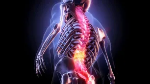
Lumbar lordosis prolonged by vertebral insertions
4. Dorso-Lumbo-Sacral Lordosis (T7-S1)
At the lumbar level, dominant vectors with direct insertion on the column are: posteriorly the paravertebral muscles, quadratus lumborum and latissimus dorsi; anteriorly the diaphragm (pillars) and iliopsoas [25]. Lumbar lordosis is prolonged by the vertebral insertions of latissimus dorsi to T7, creating a continuous functional unit [10,25].
All muscles acting with insertion on the column are co-agonists in increasing dorso-lumbar lordosis. The only antagonists are the rectus abdominis which, however, not having direct insertion on the column and being vectorially inferior, result subdominant [3,25]. The rectus abdominis have a vertical line of force that produces horizontally insignificant vectors. Their possibility of containing lumbar lordosis is linked to the capacity to stiffen the abdominal wall [26].
4.1 Biomechanical Consequences
Oblique lines of force possess vertical vectorial components which, summing with those of paravertebral muscles, increase lordosis, determine column stiffening and cause intervertebral disc compression [10,20]. Latissimus dorsi, paravertebral muscles and iliaci create a force moment that can provoke pelvic anteversion [2,10].
The equilibrium is very unstable: even modest shortening of muscles acting directly on the column determines modification of the dorso-lumbar curve [5,25]. With increased lordosis, individual G and R forces applied to each vertebral body determine, with their components, compressions on intervertebral discs. The overall G and R forces may not meet on a disc and, if their conjunction occurs on the articular facets, mechanical compressions potentially degenerative into facet fractures can be created [20,27].
4.2 Lumbar Straightening
In some radiographic pictures lumbar tract straightening is evident. Since locally muscles all act in lordosis increase, verticalization can be the resultant of extreme pelvic anteversion or dorsal kyphosis decrease [11,24].
Latissimus dorsi and paravertebral muscles bring the pelvis into anteversion. If anteversion is increased by further traction of latissimus dorsi and iliaci, in order to remain in upright station without falling forward, T7 would function as a fixed point. The lumbo-sacral curve tract will therefore be transformed into two rectilinear tracts with angular apex on the fifth lumbar vertebra [25,28]. The decrease of lumbar lordosis is therefore the product of exasperation of traction forces that determine lordosis increase, with particular involvement of the latissimus dorsi-iliaci couple.
4.3 Pelvic Equilibrium
In upright station, antero-posterior stability of the pelvis is determined by two antagonist groups [29]. For anteversion act latissimus dorsi, paravertebral muscles, iliaci and rectus femoris. For retroversion act the hamstrings and rectus abdominis. Force vectorial dominance favours anteversion, principally due to the great traction force expressible by latissimus dorsi [3,25,29].
5. Synthesis and Clinical Implications
Vector analysis of the vertebral column in the sagittal plane reveals how physiological curves result from dynamic equilibrium between dominant and subdominant muscular forces. The myofunctional subdivision into cranio-cervico-dorsal lordosis (C0-T3), dorsal kyphosis (T4-T6) and dorso-lumbo-sacral lordosis (T7-S1) reflects the real distribution of muscular insertions and consequent vectorial actions [8,11].
The identified vectorial dominances enable understanding of the most frequent patterns of postural alteration. In the cranio-cervico-dorsal tract, posterior muscles are vectorially dominant and determine lordosis increase; anterior muscles can invert their action according to cranial position [9,17]. In the dorsal kyphotic tract, scapular adductors are dominant over serratus anterior, determining hypokyphosis at apex T5 [22]. In the dorso-lumbar tract, all muscles with vertebral insertion are co-agonists in increasing lordosis, with rectus abdominis subdominant [25].
This understanding has important clinical implications. Therapeutic intervention should target muscles primarily responsible for alterations, rather than muscles expressing compensatory activity [5,14]. An electromyographically "hyperactive" muscle may simply be a compensator necessary to maintain equilibrium against the force of a shortened muscle showing low EMG activity but high passive resistant force [1,5].
Disc compressions derive not only from curve increases, but from the sum of vertical components of oblique muscular forces [10,20]. This explains why even apparently opposite pictures (hyperlordosis and straightening) can both generate disc compressions, albeit with different distribution.
Vector analysis thus provides an interpretive tool that transforms clinical observation into precise biomechanical diagnosis. Every clinical datum becomes a visible expression of physical relationships governing the entire system. Physiological curves do not represent fixed configurations, but dynamic adaptations generated by continuous balancing between G and R forces, M moments and elastic properties of tissues [2,3,10]. In this perspective, the shape of the column is not rigid anatomical data, but the result of a vectorial system in unstable equilibrium.
We hope this article was helpful. For more information from AIFiMM Formazione, please visit their CPD Member Directory page. Alternatively, you can go to the CPD Industry Hubs for more articles, courses and events relevant to your Continuing Professional Development requirements.
References
1. Lastrico M. Clinical Assessment of Muscle Shortening. The CPD Certification Service; 2025.
2. Lastrico M. Body Equilibrium - A Physical-Clinical Interpretation of Human Upright Stability. The CPD Certification Service; 2025.
3. Lastrico M. Vector Analysis in Musculoskeletal Biomechanics - Part 1: Foundations and Clinical Principles. The CPD Certification Service; 2025.
4. Lastrico M. Vector Analysis in Musculoskeletal Biomechanics - Part 2: Clinical Applications and Case Interpretation. The CPD Certification Service; 2025.
5. Sahrmann SA. Diagnosis and Treatment of Movement Impairment Syndromes. St. Louis: Mosby; 2002.
6. Knudson D. Fundamentals of Biomechanics. 2nd ed. New York: Springer; 2007.
7. Winter DA. Biomechanics and Motor Control of Human Movement. 4th ed. Hoboken: Wiley; 2009.
8. Lieber RL, Fridén J. Functional and clinical significance of skeletal muscle architecture. Muscle Nerve. 2000;23(11):1647-1666.
9. Lieber RL, Ward SR. Skeletal muscle design to meet functional demands. Philos Trans R Soc Lond B Biol Sci. 2011;366(1570):1466-1476.
10. Panjabi MM. The stabilizing system of the spine. Part I. Function, dysfunction, adaptation, and enhancement. J Spinal Disord. 1992;5(4):383-389.
11. Harrison DD, Janik TJ, Troyanovich SJ, Harrison DE, Colloca CJ. Evaluation of the assumptions used to derive an ideal normal cervical spine model. J Manipulative Physiol Ther. 1997;20(4):246-256.
12. Shumway-Cook A, Woollacott M. Motor Control: Translating Research into Clinical Practice. 4th ed. Philadelphia: Lippincott Williams & Wilkins; 2012.
13. Dubousset J. Three-dimensional analysis of the scoliotic deformity. In: Weinstein SL, editor. The Pediatric Spine: Principles and Practice. New York: Raven Press; 1994. p. 479-496.
14. Page P, Frank CC, Lardner R. Assessment and Treatment of Muscle Imbalance: The Janda Approach. Champaign: Human Kinetics; 2010.
15. Vasavada AN, Li S, Delp SL. Influence of muscle morphometry and moment arms on the moment-generating capacity of human neck muscles. Spine. 1998;23(4):412-422.
16. Mayoux-Benhamou MA, Revel M, Vallée C. Selective electromyography of dorsal neck muscles in humans. Exp Brain Res. 1997;113(2):353-360.
17. Conley MS, Meyer RA, Bloomberg JJ, Feeback DL, Dudley GA. Noninvasive analysis of human neck muscle function. Spine. 1995;20(23):2505-2512.
18. Keshner EA. Head-trunk coordination during linear anterior-posterior translations. J Neurophysiol. 2003;89(4):1891-1901.
19. Panjabi MM, Crisco JJ, Vasavada A, Oda T, Cholewicki J, Nibu K, Shin E. Mechanical properties of the human cervical spine as shown by three-dimensional load-displacement curves. Spine. 2001;26(24):2692-2700.
20. Adams MA, Roughley PJ. What is intervertebral disc degeneration, and what causes it? Spine. 2006;31(18):2151-2161.
21. Cholewicki J, McGill SM. Mechanical stability of the in vivo lumbar spine: implications for injury and chronic low back pain. Clin Biomech. 1996;11(1):1-15.
22. Cools AM, Declercq GA, Cambier DC, Mahieu NN, Witvrouw EE. Trapezius activity and intramuscular balance during isokinetic exercise in overhead athletes with impingement symptoms. Scand J Med Sci Sports. 2007;17(1):25-33.
23. Lee LJ, Coppieters MW, Hodges PW. Differential activation of the thoracic multifidus and longissimus thoracis during trunk rotation. Spine. 2005;30(8):870-876.
24. Kado DM, Huang MH, Karlamangla AS, Barrett-Connor E, Greendale GA. Hyperkyphotic posture predicts mortality in older community-dwelling men and women: a prospective study. J Am Geriatr Soc. 2004;52(10):1662-1667.
25. McGill SM. Low Back Disorders: Evidence-Based Prevention and Rehabilitation. 3rd ed. Champaign: Human Kinetics; 2016.
26. Hodges PW, Richardson CA. Contraction of the abdominal muscles associated with movement of the lower limb. Phys Ther. 1997;77(2):132-142.
27. Adams MA, Hutton WC. The effect of posture on the role of the apophysial joints in resisting intervertebral compressive forces. J Bone Joint Surg Br. 1980;62(3):358-362.
28. Roussouly P, Gollogly S, Berthonnaud E, Dimnet J. Classification of the normal variation in the sagittal alignment of the human lumbar spine and pelvis in the standing position. Spine. 2005;30(3):346-353.
29. Barker PJ, Guggenheimer KT, Grkovic I, Briggs CA, Jones DC, Thomas CD, Hodges PW. Effects of tensioning the lumbar fasciae on segmental stiffness during flexion and extension: Young Investigator Award winner. Spine. 2006;31(4):397-405.